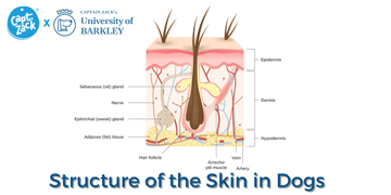
The skin is the largest organ of your dog’s body. It provides a protective barrier against the environment, regulates temperature, and gives your dog its sense of touch. Depending on the species and age, the skin may be 12 to 24% of a dog’s body weight. The skin has 3 major layers: the epidermis or outermost layer, the dermis or middle layer, and subcutis or innermost layer. Other important parts of the skin include skin appendages (such as hair and claws) and subcutaneous muscles and fat.
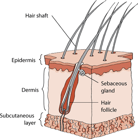
The anatomy of a dog’s skin includes 3 major layers, as well as hair follicles and sebaceous glands.
|
Epidermis
The epidermis is the outer layer of skin. It provides protection from foreign substances. The epidermis is composed of multiple types of cells, including keratinocytes, melanocytes, Langerhans cells, and Merkel cells. Each of these cells has special functions.
Keratinocytes provide a protective layer that is constantly being renewed in a process called keratinization. In this process, new skin cells are created near the base of the epidermis and migrate upwards. This produces a compact layer of dead cells on the skin surface. This layer keeps in fluids, salts, and nutrients, while keeping out infectious or noxious agents. The top layer of dead skin cells are continuously shed and replaced by cells from lower layers. The rate of cell replacement is affected by nutrition, hormones, tissue factors, immune cells in the skin, and genetics. Disease, some drugs, and inflammation also alter normal cell growth and keratinization.
Melanocytes are located at the base of the epidermis, the outer root sheath of hairs, and the ducts of the oil and sweat glands. The melanocytes produce the skin and hair coloring (pigment) called melanin. Production of melanin is controlled by both hormones and the genes received from parents. Melanin helps protect the cells from the damaging rays of the sun.
Langerhans cells are part of the immune system. These cells are damaged when exposed to excessive ultraviolet light and glucocorticoids (anti-inflammatory drugs). Langerhans cells play an important role in the skin’s response to foreign substances and contribute to such things as the development of rashes when an animal is exposed to irritating materials.
Merkel cells are specialized cells associated with the sensory organs in the skin. In particular, Merkel cells help provide animals with sensory information from whiskers and the deep skin areas called tylotrich pads.
Basement Membrane Zone
This layer of the skin is located beneath the epidermis and connects the epidermis to the dermis layer below. It also serves as a protective barrier between the epidermis and the dermis. Several skin diseases, including a number of autoimmune conditions, can damage the basement membrane zone.
Dermis
The dermis supports and nourishes the epidermis and skin appendages. The blood vessels that supply the epidermis with nutrients are located in the dermis. Blood vessels also regulate skin and body temperature. Sensory nerves are located in the dermis and hair follicles. Motor nerves are also present. The skin responds to the sensations of touch, pain, itch, heat, and cold. The dermis secretes the protein collagen, which supports the skin. There are also immune cells in the dermis that defend against infectious agents that pass through the epidermis.
Skin Appendages
Hair follicles, oil and sweat glands, and claws are skin appendages that grow out of the epidermis. The hair follicles of dogs are compound, which means the follicles have a central hair surrounded by 3 to 15 smaller secondary hairs all exiting from one pore. Dogs are born with simple hair follicles that develop into compound hair follicles.
The growth of hair is affected by nutrition, hormones, and change of season. Dogs normally shed hair in the early spring and early fall. They may also shed in response to changes in temperature or amount of sunlight. The size, shape, and length of hair are controlled by genetics. Hormones, disease, drugs, nutrition, and environment also affect the health of hair.
The hair coat protects the skin from physical and ultraviolet light damage, and helps regulate body temperature. Trapping air between secondary hairs conserves heat. This requires that the hairs be dry and waterproof. The cold-weather coat of many dogs is longer and finer to facilitate heat conservation. The hair coat can also help cool the skin. The warm-weather coat has shorter, thicker hairs and fewer secondary hairs. This anatomic change allows air to move easily through the coat, which facilitates cooling. Hair coats can also act as camouflage to conceal wild animals.
Oil glands (also called sebaceous glands) secrete an oily substance called sebum into the hair follicles and onto the skin. They are present in large numbers near the paws, back of the neck, rump, chin, and tail area. Sebum is a mixture of fatty acids. Sebum is important for keeping the skin soft, moist, and pliable. Sebum gives the hair coat sheen and has antibiotic properties.
Dogs have sweat glands on the feet that may have a minor role in cooling of the body. However, dogs primarily release excess body heat by panting and drooling.
Subcutis
The subcutis is the innermost layer of the skin. It contains the subcutaneous fat and muscles. (The word subcutaneous means “beneath the skin.”) The twitch muscle is the major muscle immediately beneath the skin. The subcutaneous fat provides many functions, including insulation; a reservoir for fluids, electrolytes, and energy; and a shock absorber.